Models
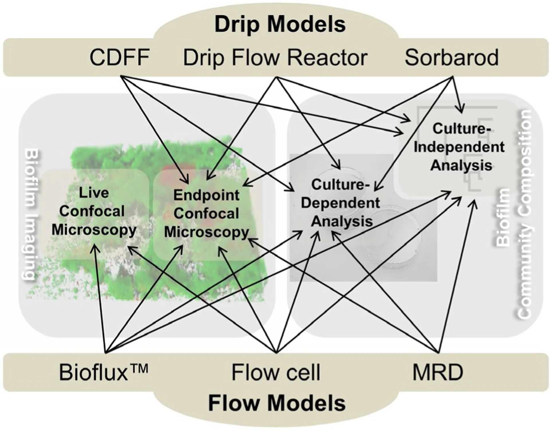
A key focus of the Rickard lab is to use in vitro models to investigate cell-cell interactions and biofilm development that occur in natural environments. To achieve this, standard laboratory biofilm models (e.g. microplate-based models) through more advanced models (e.g. flowcells and microfluidic systems) are used to examine interactions that promote the development of single or multi-species biofilms and explore ways to control them (e.g. antimicrobial treatments).
The model systems we use are (1) often compatible with culture and culture independent techniques to evaluate biofilm biomass/species composition and (2) amenable to confocal laser scanning microscopy to visualize and perform image analysis of the communities.
SPH XR Lab Safety Training
In partnership with the U-M Department of Environmental Health and Safety and SPH Media Services, efforts are underway to enhance laboratory health and safety training for new researchers. By integrating Extended Reality (XR) technology, the initiative to boost engagement and improve retention of essential lab safety practices—transforming what is traditionally delivered through text-based materials into an immersive, interactive learning experience.

Collaborators

Dave Bridges, PHD | XR

Chris Fenno, PHD | Oral Biofilms

Dustin Moore | XR

Nicholas Jakubovics, PHD | Biofilms

John Cessna | XR

Livia Tenuta, DDS, PHD | Oral Biofilms
Not Pictured: Meg Vickerman, PHD

Eric Grzyb | XR